
Degenerative Disc Disease in Greensburg, PA
The name frightens people more than the finding warrants. Degenerative change is common with age and is frequently present without symptoms. We measure what is actually relevant to your presentation and explain honestly what care can and cannot influence.
Schedule a Complimentary Structural ConsultationStart With the Name
“Degenerative disc disease” is a misleading label, and it does real harm. It is not a disease in the usual sense — it describes age- and load-associated changes in the discs: loss of water content, reduced disc height, and the bony changes that accompany them.
A systematic review of spinal imaging in people with no symptoms at all found disc degeneration in a substantial share of people in their twenties, rising to nearly everyone by their eighties.1 If a finding is present in most healthy, pain-free people of a given age, it cannot by itself be the explanation for one person’s pain.
Many patients arrive having been shown a scan and told their spine is “wearing out” or that they will end up disabled. That framing is not supported by the evidence, and the fear it creates can make pain and disability worse. We will look at the actual findings with you.
Why Imaging Severity and Symptoms Disagree
This is the single most useful thing to understand about this diagnosis. Two people can have nearly identical imaging and completely different experiences: one in significant pain, one entirely comfortable. Conversely, significant pain often occurs with unremarkable imaging.
Pain is influenced by mechanical loading, movement, conditioning, sleep, stress, and sensitisation — not by disc height alone. This is why we assess the person and the mechanics, not the report.
Why One Segment More Than Another?
Age explains why discs change. It explains less well why one level shows marked change while the level directly above looks comparatively healthy. Segments are not loaded equally, and the factors that influence distribution include:
- • Spinal alignment and how load is shared across segments
- • Previous injury to a particular level
- • Mobility or stiffness of adjacent segments
- • Individual anatomy and genetics
- • Occupational and cumulative loading history
Alignment is the factor we can measure objectively, and where our examination identifies a Structural Shift it is worth addressing. It is one contributor among several — not the sole cause of degeneration, and we would not claim otherwise.
A Documented Lumbar Curve Correction
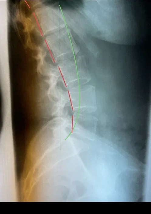
Reduced lumbar curve
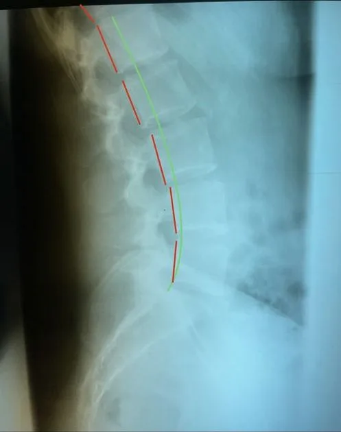
Improved lumbar curve
Improving the lumbar curve changes how load is distributed across the segments. This shows a change in alignment — not regeneration of disc tissue.
An individual patient result. Individual results vary, and a clinical evaluation is required to determine candidacy.
What Else Can Feel Similar?
Because degenerative findings are so common, they are frequently blamed for symptoms arising elsewhere:
- • Facet joint pain, which often refers into the buttock and thigh
- • Sacroiliac joint pain
- • Spinal stenosis, especially where walking provokes leg symptoms
- • Disc herniation with genuine nerve-root involvement
- • Hip pathology masquerading as spinal pain
- • Muscular and myofascial referral
- • Inflammatory arthropathy, which behaves differently and needs medical diagnosis
When to Seek Urgent Medical Evaluation
Degenerative change itself is not an emergency, but certain neurological or systemic findings require prompt evaluation. Seek immediate emergency evaluation if you develop:
- • New difficulty controlling your bladder or bowels
- • Numbness in the saddle or groin area
- • Weakness that is severe, worsening, or affecting both legs
- • Rapidly progressing numbness or loss of function
Together these can indicate cauda equina syndrome.2 Separately, back pain accompanied by fever, unexplained weight loss, pain that consistently wakes you at night, or a history of cancer should be assessed medically before structural care is considered — degenerative change on a scan does not rule those out.
How We Evaluate It
- Consultation and history — what provokes and relieves symptoms, and how they have behaved over time.
- Neurological examination — whether a nerve root is genuinely involved.
- Orthopedic and functional testing — movement, load tolerance, and which structures reproduce your symptoms.
- Posture and structural analysis — measuring how load is distributed, which is the part we can actually influence.
- Existing imaging in context — if you have a report describing degeneration, we will tell you honestly how much of it is age-expected.
When Imaging May Be Appropriate
Routine imaging is not recommended for uncomplicated back pain, partly because degenerative findings are near-universal with age and frequently mislead.3 Where imaging is indicated: structural X-ray shows alignment, disc-space height, and bony change; MRI shows the disc, nerve roots, and canal directly and is preferred when neurological involvement needs to be assessed.
What Care May Include
- • Reducing mechanical loading on the affected segment
- • Specific adjusting directed at measured structural findings
- • Traction strategies in selected cases where clinically appropriate
- • Rehabilitative exercise to build load tolerance — the most durable factor in most cases
- • Reassurance and accurate explanation, which genuinely matters here
What we do not claim.
We do not claim to reverse degeneration, regenerate disc tissue, restore lost disc height, or halt the progression of age-related change. We also do not claim that traction pumps nutrients back into a disc. What we can measure is alignment, examination findings, and your function — and those are what we report back to you.
Who May Be an Appropriate Candidate
People with mechanical or activity-related symptoms, no identified red flags, and examination findings reasonably consistent with their presentation may be appropriate candidates for conservative care. The decision depends on the individual examination, neurological findings, imaging when indicated, and the patient’s goals.
When Another Evaluation May Be More Appropriate
Medical evaluation, co-management, or referral may be appropriate when examination findings suggest a condition outside the scope of conservative musculoskeletal care, when significant or progressive neurological deficits are present, when serious pathology is suspected, or when the presentation requires diagnostic evaluation or treatment beyond what can appropriately be provided in this office.
Referral is not the same as refusal. Many of these situations simply mean another opinion comes first, or that care is shared — we will explain which applies to you.
How Progress Is Measured
- • Change in symptom intensity, frequency, and pattern
- • Function — standing and sitting tolerance, walking, lifting, work capacity
- • Movement and load tolerance on re-examination
- • Repeat posture or alignment measurement where those findings were relevant
- • Repeat neurological findings where a deficit was present initially
We do not re-image to look for “less degeneration”, because that is not a realistic or meaningful measure of whether care is working.
What People Commonly Try
Most people arrive having already tried at least one of these. Each has a legitimate place, and for many people with degenerative disc findings one of them is the right answer. The question is whether anything structural has been measured along the way.
Activity modification and rest
Where it helps: Often the right first step. Easing the activities that provoke symptoms can settle a flare-up and is sensible early on.
Its limit: It says little about what is driving the problem, and prolonged rest tends to reduce tolerance rather than build it.
Over-the-counter or prescribed medication
Where it helps: Can reduce pain and inflammation enough to keep you moving and sleeping, which matters. Its appropriate use is a conversation with your physician.
Its limit: It is aimed at the symptom rather than at any structural or mechanical finding, so it does not change what is measured.
Exercise and stretching
Where it helps: Among the most strongly supported options for many spinal conditions, and part of most good plans, including ours.
Its limit: A general programme is not matched to measured findings, so if a structural finding is present it can strengthen around it without addressing it.
Physical therapy
Where it helps: Strengthening, mobility and movement retraining with a qualified professional. Many people do well with it.
Its limit: Whether structural alignment is measured and re-measured varies by provider; progress is often judged mainly by symptoms and function.
Injections
Where it helps: Can calm inflammation around an irritated nerve or joint, sometimes enough to allow other care to proceed.
Its limit: The effect is usually temporary, repeat use is limited, and the structure is unchanged afterwards.
Chiropractic care aimed at relief
Where it helps: Often effective for its goal of reducing pain and improving how you move, and sometimes all a person needs.
Its limit: Progress is usually judged by symptoms, and structural measurement is not always part of the process.
None of this is an argument against those options. It is the reason we measure before recommending anything. How relief-focused care and structural correction differ.
How Optimus Approaches the Evaluation
The process is the same for degenerative disc findings as for any presentation we see, because it is designed to find out whether structure is part of your problem rather than to assume it is.
Complimentary Structural Consultation
A private conversation about your concerns, goals, previous care and whether this office is an appropriate fit. No examination, imaging or treatment takes place.
Paid structural examination
Orthopedic, neurological, movement and functional testing. The fee is explained before it is scheduled.
Objective measurements
Posture measured in millimeters and compared with normal reference values, so there is a baseline to re-measure against.
Imaging only when clinically warranted
Structural X-rays or stress views are taken only when the examination indicates them, and the reason is explained first.
Patient Recommendation Conference
The separate visit where your findings, our recommendations, the proposed plan and the fees are explained before you agree to anything.
Individualized recommendations
If structural correction is appropriate, a plan built around your own measured findings. If another evaluation is the better path, we say so.
Scheduled progress examinations
Re-examination at set points so change is documented, not assumed. If the measurements are not changing, the approach changes.
Comparative imaging only when clinically justified
Repeat images are taken to compare against the baseline only when there is a clinical reason to do so.
What the first visits involve, what to bring and how payment works are set out on the new patient page.
Not Sure Whether This Applies to You?
The complimentary structural consultation is the place to ask. It is a private conversation about your degenerative disc findings, what you have already tried and whether measuring your spine is a reasonable next step. No examination, imaging or treatment takes place, nothing is booked beyond it, and if another kind of evaluation is the better path we will say so.
Schedule a Complimentary Structural ConsultationFrequently Asked Questions
Is degenerative disc disease actually a disease?
Not in the way the name suggests, and the terminology causes real distress. It describes age- and load-associated changes in the discs — loss of hydration, reduced disc height, and related bony changes. These are found in the majority of people as they age, including people with no back pain whatsoever. It is closer to grey hair than to a progressive illness.
Does it get worse over time?
Disc changes generally accumulate with age, but that is not the same as symptoms getting worse. Many people's pain improves or fluctuates while their imaging stays the same or shows further change. Imaging severity and symptom severity correlate poorly, which is why we do not treat the report.
Will I end up disabled or in a wheelchair?
That is a fear many patients arrive with, often after being shown a scan without much context. Degenerative change on imaging is extremely common and, on its own, is not a prediction of disability. If you have been told otherwise, we are happy to look at the actual findings with you and explain what they do and do not indicate.
Can degeneration be reversed?
No, and we will not claim otherwise. Established structural change in a disc — lost height, altered composition, bony change — is not something conservative care reverses. What can often be influenced is the mechanical loading on the segment, movement and load tolerance, and symptoms. Those are what we measure.
Why does one disc degenerate more than the one above it?
Segments are not loaded equally. Alignment, previous injury, mobility of adjacent segments, and individual anatomy all influence which levels take more mechanical stress. That is one reason we measure alignment — it is a factor we can actually assess and, where relevant, address.
Do I need an X-ray or MRI?
Not automatically. Degenerative change is usually visible on X-ray, which is useful for alignment and disc-space height. MRI shows the disc itself and the nerve roots. But because these findings are so common with age, imaging often does not change what should be done next in an uncomplicated presentation.
When should I seek urgent evaluation?
New difficulty controlling your bladder or bowels, numbness in the saddle or groin area, or weakness that is severe or worsening warrants immediate emergency assessment. Back pain with fever, unexplained weight loss, night pain that consistently wakes you, or a history of cancer should be evaluated medically before structural care.
Research and References
Cited for background on the prevalence of degenerative findings, imaging indications, and emergency red flags — not as proof of any chiropractic outcome.
- 1 Brinjikji W, Luetmer PH, Comstock B, et al. Systematic literature review of imaging features of spinal degeneration in asymptomatic populations. AJNR Am J Neuroradiol. 2015;36(4):811–816. PMID 25430861
- 2 Rider LS, Marra EM. Cauda Equina and Conus Medullaris Syndromes. StatPearls [Internet]. StatPearls Publishing. PMID 30725885
- 3 Qaseem A, Wilt TJ, McLean RM, Forciea MA, et al. Noninvasive Treatments for Acute, Subacute, and Chronic Low Back Pain: A Clinical Practice Guideline From the American College of Physicians. Ann Intern Med. 2017;166(7):514–530. PMID 28192789
Related Reading
Published by Optimus Spine & Posture, Greensburg, PA. Learn more about Dr. Gareth Bury, DC.
Individual results vary. A clinical evaluation is required to determine candidacy for structural correction. This page is educational and is not a substitute for medical evaluation.
Common Symptoms
- • Back or neck pain that varies day to day
- • Stiffness, often worse after rest
- • Symptoms eased by movement or position change
- • Referred pain into the buttock or shoulder
- • Numbness or weakness if a nerve is involved
Most people with degenerative findings on imaging have no symptoms at all.
Find Out What Your Scan Actually Means
If you have been told your spine is “wearing out”, it is worth understanding how much of that is age-expected and how much is relevant to you.
Your initial structural consultation is complimentary and comes with no obligation. It does not include the comprehensive examination, diagnostic imaging or treatment, which are paid services — and imaging is performed only when it is clinically warranted. When appropriate, the consultation and examination may occur during the same visit. Fees are explained before you agree to anything.